
|
|
VIII. Answer the following questions. Use all information given before.1. What enables a multicellular organism to grow and replace worn out or damaged cells? 2. What does cell division start with? 3. What two forms of nuclear division do you know? 4. What are chromosomes? 5. What do chromosomes consist of? 6. What is known as homologous pairs? 7. What is the difference between diploid and haploid? 8. What are three main stages of the cell cycle? 9. How does the duration of the cell cycle vary? IX. Match the sentence halves. Make complete sentences:
X. Read and translate the short text without any dictionary: Fact of life: Some laboratory-grown mammalian cells appear to obey an internal “biological clock” that allows them to divide by mitosis a maximum number of times. For example, a fibroblast (connective tissue cell) taken from a fetus divides on average about 50 times; the same type of cell taken from an adult divides only 14 to 19 times.
XI. Food for thought:Although meiosisoccurs at some stage in the life cycle of sexually reproducing plants, their gametes are usually formed by mitosis. Suggest reasons for this. Text 2.3. Microscopes
■ Essential targets: By the end of this text you should be able to: ● describe the main features of a light microscope and an electron microscope ● distinguish between the terms magnification and resolving power ● give the approximate size of different biological structures using an appropriate unit of measurement. Pre-reading ■ Discuss these questions with your partner. 1.Who invented a microscope? 2. What types of microscopes are used today?
■ Read the given text and make your essential assignments:
The compound light microscope The compound light microscope is also called a light microscope or optical microscope. The compound light microscope is also called a light microscope or optical microscope. The lenses refract (bend) the light to give a magnified image of the object. The image may be projected directly into the viewer’s eye or into photographic film. A photograph taken through a light microscope is called a photomicrographor light micrograph. Magnification and resolution The magnificationof an instrument is the increase in the apparent size of the object. The total magnification of a compound microscope is worked out by multiplying the magnification of the objective lens by that of the ocular lens. There is virtually no limit to the magnification produced by a light microscope; it depends on the power of the lenses used. However, above a certain magnification the image becomes blurred and it is impossible to distinguish structures lying close together. This limit of effective magnification is called the resolving poweror resolution of the microscope. It is defined as the ability of a microscope to show two objects as separate. The resolving power of the light microscope is limited by the wavelength of light. Light microscopes can magnify objects up to about 1500 times without losing clarity. The electron microscope Electron microscopes use a beam of electrons instead of a beam of light. Electron beams have a much smaller wavelength than light rays, so electron microscopes have greater resolving powers and can produce much higher effective magnifications than light microscopes. There are two main types of electron microscopes: thetransmission electron microscope(TEM), and the scanning electron microscope(SEM). 
Не нашли, что искали? Воспользуйтесь поиском по сайту: ©2015 - 2024 stydopedia.ru Все материалы защищены законодательством РФ.
|

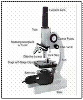 A microscope is used to produce a magnified image of an object or specimen. Anton van Leeuwenhoek (1632-1723) was the first to invent a microscope powerful enough to explore the world of microbes. His discoveries stimulated an explosion of interest in the scientific use of microscopes. Since the 18th century many new types have been invented, of which the most commonly used today are the compound light microscopeand the electron microscope.